Documents
Poster
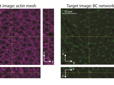
PREDICTION OF MULTIPLE 3D TISSUE STRUCTURES BASED ON SINGLE-MARKER IMAGES USING CONVOLUTIONAL NEURAL NETWORKS
- Citation Author(s):
- Submitted by:
- Hernan Morales-...
- Last updated:
- 20 September 2019 - 7:25am
- Document Type:
- Poster
- Document Year:
- 2019
- Event:
- Presenters:
- Hernan Morales-Navarrete
- Paper Code:
- 2314
- Categories:
- Keywords:
- Log in to post comments
A quantitative understanding of complex biological systems such as tissues requires reconstructing the structure of the different components of the system. Fluorescence microscopy provides the means to visualize simultaneously several tissue components. However, it can be time consuming and is limited by the number of fluorescent markers that can be used. In this study, we describe a toolbox of algorithms based on convolutional neural networks for the prediction of 3D tissue structures by learning features embedded within single-marker images. As proof of principle, we aimed to predict the network of bile canaliculi (BC) in liver tissue using images of the cortical actin mesh as input. The actin meshwork has a characteristic organization in specific cellular domains, such as BC. However, the use of manually selected features from images of actin is not sufficient to properly reconstruct BC structure. Our deep learning framework showed a remarkable accuracy for the prediction of BC network and was successfully adapted (i.e. transfer learning) to predict the sinusoidal network. This approach allows for a complete reconstruction of tissue microarchitecture using a single fluorescent marker.

