- Read more about ADABOOST-BASED DETECTION AND SEGMENTATION OF BIORESORBABLE VASCULAR SCAFFOLDS STRUTS IN IVOCT IMAGES
- Log in to post comments
In this paper, we presented an automatic method for BVS struts detection in IVOCT images based on Haar-like features and Adaboost algorithm. Then, DP algorithm was used for struts segmentation. Based on the detection and segmentation results, apposed and malapposed struts were distinguished automatically. The qualitative and quantitative evaluation shows that our method is effective and robust for BVS struts detection and segmentation, and is capable of malapposition analysis.
ICIP_POSTER2501.pdf
- Categories:
 11 Views
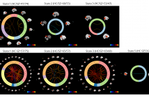
- Read more about Identifying fMRI Dynamic Connectivity States Using Affinity Propagation Clustering Method: Application to Schizophrenia
- Log in to post comments
Numerous studies have shown that brain functional connectivity patterns can be time-varying over periods of tens of seconds. K-means has been widely used to extract the connectivity states from dynamic functional connectivity. However, K-means is dependent on initialization and can be exponentially slow in converging due to extensive noise in dynamic functional connectivity. In this work, we propose to use an affinity propagation clustering method to estimate the connectivity states. The new approach found more meaningful group differences than K-means. Our finding supports that our method is promising in exploring biomarkers of mental disorders.
- Categories:
9 Views- Read more about Classification of Thyroid Nodules in Ultrasound Images Using Deep Model Based Transfer Learning and Hybrid Features
- Log in to post comments
Ultrasonography is a valuable diagnosis method for thyroid nodules. Automatically discriminating benign and malignant nodules in the ultrasound images can provide aided diagnosis suggestions, or increase the diagnosis accuracy when lack of experts. The core problem in this issue is how to capture appropriate features for this specific task. Here, we propose a feature extraction method for ultrasound images based on the convolution neural networks (CNNs), try to introduce more meaningful semantic features to the classification.
- Categories:
46 ViewsBiomedical images are usually corrupted by strong noise and
intensity inhomogeneity simultaneously. Existing regionbased active contour models (RACMs) easily fail when segmenting such images. In the frequency domain, we propose a
generalized RACM that presents a new way to understand the
essence of classical RACMs whose segmentation results are
determined by a frequency filter to extract the proposed frequency boundary energy. Then, we introduce the difference
of Gaussians as the optimal filter to exclude strong noise and
- Categories:
13 Views- Read more about Automatic segmentation of retinal vasculature
- Log in to post comments
- Categories:
4 Views
- Read more about A SEMI-SUPERVISED METHOD FOR MULTI-SUBJECT FMRI FUNCTIONAL ALIGNMENT
- Log in to post comments
Practical limitations on the duration of individual fMRI scans have led neuroscientist to consider the aggregation of data from multiple subjects. Differences in anatomical structures and functional topographies of brains require aligning data across subjects. Existing functional alignment methods serve as a preprocessing step that allows subsequent statistical methods to learn from the aggregated multi-subject data. Despite their success, current alignment methods do not leverage the labeled data used in the subsequent methods.
- Categories:
9 Views- Read more about Active Learning for Magnetic Resonance Image Quality Assessment
- Log in to post comments
In medical imaging, the acquired images are usually analyzed by a human observer and rated with respect to a diagnostic question. However, this procedure is time-demanding and expensive. Furthermore, the lack of a reference image makes this task challenging. In order to support the human observer in assessing image quality and to ensure an objective evaluation, we extend in this paper our previous no-reference magnetic resonance (MR) image quality assessment system with an active learning loop to reduce the amount of necessary labeled training data.
- Categories:
28 Views- Read more about Implicit kernel presentation aware object segmentation framework
- Log in to post comments
Given a set of training shapes and an input image with a shape similar to some of the elements in the training set, this poster introduces a new implicit kernel sparse model with a twofold goal. First, to obtain an implicit kernel sparse neighbor based combination that best represents the object. Second, to accurately segment the object taking into accounts both the high-level implicit kernel presentation and the low-level image information.
- Categories:
8 Views- Read more about A novel array processing method for precise depth detection of ultrasound point scatter
- Log in to post comments
A signal based algorithm resulting in increased depth resolution is presented for medical ultrasound. It relies on multiple foci beamforming that is enabled by current ultrasound imaging systems. The concept stems from optical microscopy and is translated here into ultrasound using the Field II simulation software. A 7 MHz linear transducer is used to scan a single point scatterer phantom that can move in the axial direction.
- Categories:
7 Views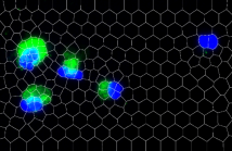
- Read more about MULTI-RESOLUTION SUPER-PIXELS AND THEIR APPLICATIONS ON FLUORESCENT MESENCHYMAL STEM CELLS IMAGES USING 1-D SIFT BASED MERGING
- Log in to post comments
- Categories:
12 Views